
Lab Services > FIB Micromachining Laboratory
Imaging & Analysis
FIB Milling Capability Sample Applications Imaging & Analysis
The variety of imaging and analysis capabilities in Oregon Physics' focused ion beam (FIB) micromachining laboratory allows us to capture information of interest to you, our customer.
- We can take secondary electron and ion images with the liquid metal ion source (LMIS) FIB or with the plasma FIB.
- We have an in-house Amray 1280 scanning electron microscope (SEM) for medium resolution imaging and metrology.
- We have access to Field Emission SEMs for high resolution backscattered and secondary electron imaging.
- We have EDAX material analysis capabilities.
Examples
 |
 |
|
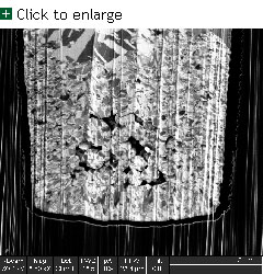
A Gallium LMIS FIB image of a cross-sectioned copper via showing large contrast between the copper grains. |
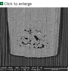
A backscattered electron image from a scanning electron microscope (SEM) showing the same cross-sectioned copper via. In this image there is good contrast showing small voids in the copper via. |
Oregon Physics, LLC
19075 NW Tanasbourne Dr., Suite 150
Hillsboro, OR 97124 USA
Phone: 503.601.0041 Fax: 503-992-6710 Email: info@oregon-physics.com
Web Design by Rareheron Web Design, Portland, OR